Symptomology
Alexander disease is an astrocytopathy that presents as a leukodystrophy in young children and a progressive neurodegenerative disorder in adults.2,5-8 Symptoms vary significantly depending on age of onset.1,4,9,10 Many pediatricians and general neurologists may not be familiar with the characteristic features of Alexander disease due to the rare nature of the condition.1,11 However, with raised awareness and education, the clinical constellation of symptoms and MRI features of Alexander disease can be recognized.
![A man with a beard lies on his back about to enter an MRI machine. [66]](/sites/default/files/2025-05/Mask%20Group%204.png)
![Four stock black and white photos appear in equal squares, from left to right: a man entering an MRI machine, an infant girl held and holding hands with her mother, a boy leaning on his father’s back, and a young girl lying on an adult’s chest. [245]](/sites/default/files/2025-05/Mask%20Group%2010.png)


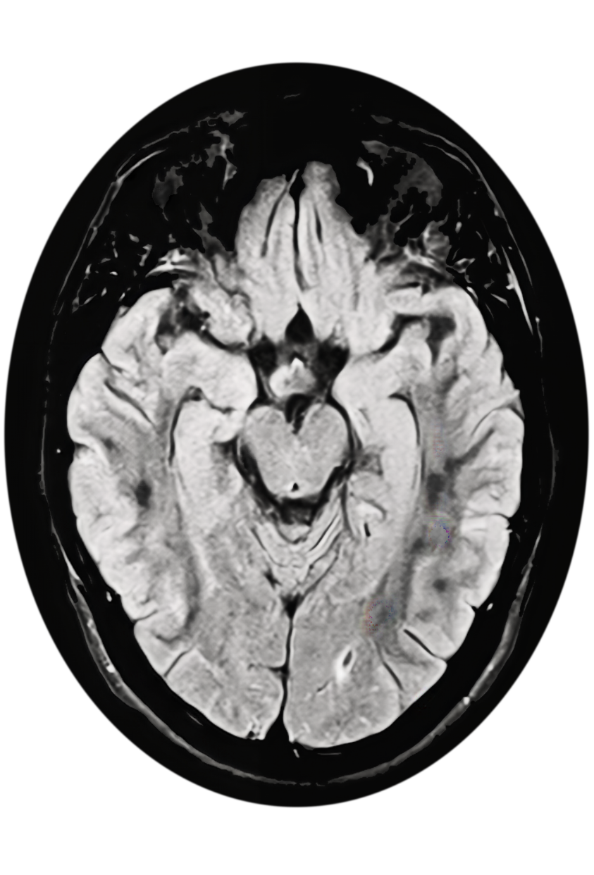
MRI features
When a leukodystrophy or Alexander disease is suspected, prompt referral to a neurologist for a brain MRI and genetic testing is critical for timely diagnosis.10-13 Neuroimaging findings of frontally predominant white matter abnormalities, consistent with a leukodystrophy, are observed in those with earlier age of onset.1,15 In those with a later age of onset, neurodegenerative features with brainstem lesions and/or atrophy of the brainstem, cerebellum, and/or cervical spinal cord are typically observed.4,14,16
Standard of care
Current approaches to disease management can mitigate symptoms but do not address the underlying cause of disease1,8,17; however, ongoing research and a clinical trial are exploring potential disease-modifying therapies.18,19