Brain MRI
EARLIER AGE OF ONSET
EARLIER AGE OF ONSET
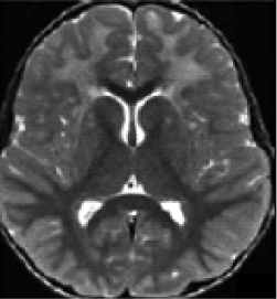
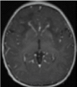
A radiologic diagnosis of Alexander disease is based on the presence of 4 out of 5 MRI features, which are most commonly observed in people with earlier age of onset (ie, infants and young children)1,15,16:
- Frontally predominant white matter changes1
- Periventricular rim abnormalities (eg, high T1-weighted signal and low T2-weighted signal)1
- Abnormalities of the basal ganglia and thalami1
- Brainstem abnormalities1
- Contrast enhancement of 1 or more of the following structures: ventricular lining, frontal white matter, optic chiasm, basal ganglia, thalamus, fornix, dentate nucleus, and brainstem1
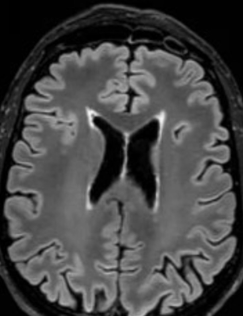
Frontally predominant cerebral white matter abnormalities14
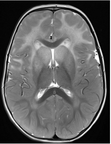
Signal abnormality and swelling (and subsequent atrophy) of the basal ganglia and thalami14
Periventricular rim of low signal intensity on T2-weighted images and high signal intensity on T1-weighted images14
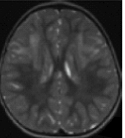
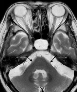
Areas of signal abnormality in the posterior part of the midbrain and the central part of the medulla14
Contrast enhancement involving ventricular lining, periventricular rim, frontal white matter, optic chiasm, fornix, basal ganglia, or thalami14
Later age of onset
The MRI features of Alexander disease in people with later age of onset (ie, juveniles and adults) do not typically include frontal white matter changes and are instead characterized by brainstem, cerebellum, and spinal cord abnormalities; these patients may not exhibit the same MRI features that support a diagnosis in people with earlier age of onset.4,16,51
MRI findings in people with onset in adulthood are characterized by:
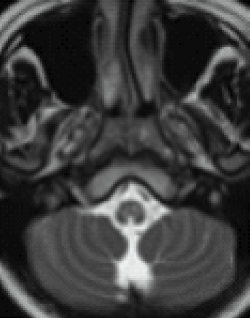
- Atrophy and changes in signal intensity in the cerebellum, medulla oblongata, and upper spinal cord, with variable supratentorial periventricular white matter abnormalities4
- A pattern of prominent atrophy involving the medulla oblongata and cervical spinal cord with sparing of the lower pons (ie, “tadpole sign”)8,52
Unlike multiple sclerosis, Alexander disease is not associated with generalized brain atrophy23; it may also be distinguished by the presence of symmetrical lesions on MRI.53 A spectrum of atypical MRI findings, including ventricular garlands, swelling of the medulla, tumor-like presentations, and inflammatory features, have also been documented.54-60
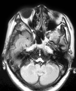
An additional imaging pattern characterized by bilateral symmetric hyperintensity on T2-weighted imaging involving the central inferior olivary nucleus, the pyramids, and the cuneate and gracile fascicles, with sparing of the peripheral inferior olivary nucleus and inferior peduncle (termed the “chipmunk sign”), is a common feature in people with Alexander disease regardless of age of onset.61
Moderate supratentorial periventricular white matter abnormalities
Atrophy of the cerebellum and middle cerebellar peduncles
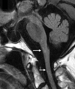
Mild atrophy of the medulla oblongata and upper cervical cord with preservation of the pons
Severe atrophy of the medulla oblongata, cerebellum, and midbrain with preservation of the pons
Images from Lynch et al. Forthcoming 2025.
Alexander disease should be considered in a differential diagnosis of people who present with suggestive clinical symptoms and MRI findings.1